It takes two: Analyzing Neural Activity From Calcium Imaging
By Sara Vaccar
Media InquiriesA growing body of researchers are utilizing optical imaging to monitor activity in the brain. One type of optical imaging, two-photon calcium imaging, has been widely adopted for its ability to record large neural populations. A major question outstanding, though, is what statistical methods to use to analyze calcium imaging recordings.
“Prior to our work, two standard types of statistical methods were applied to calcium imaging recordings—deconvolution and dimensionality reduction,” explained Steve Chase, professor of biomedical engineering at Carnegie Mellon and the Neuroscience Institute. “After testing each technique using a collection of simulated and laboratory data, we concluded that it didn’t make sense to do either/or. Combining the approaches into one operation proved to be more successful.”
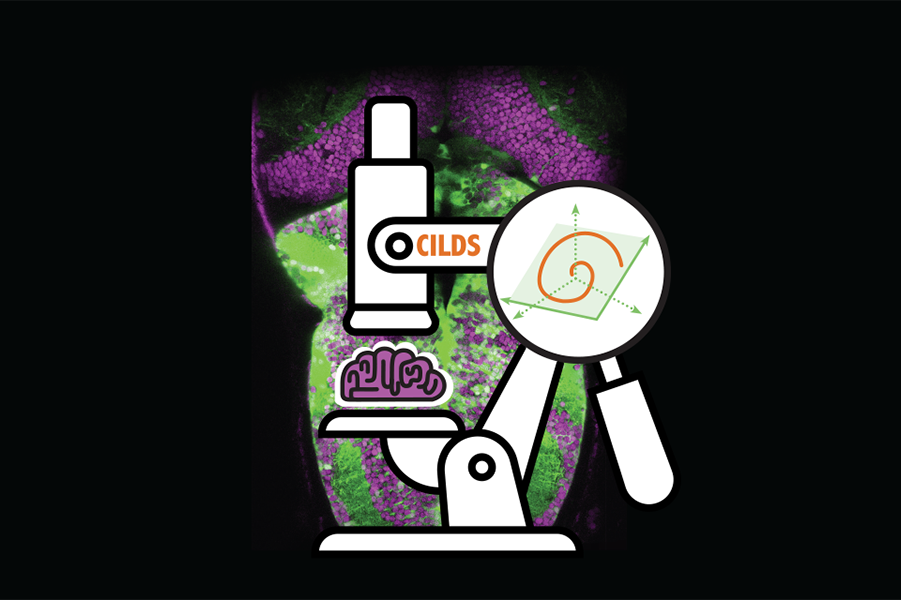
Carnegie Mellon University and Howard Hughes Medical Institute researchers teamed up to analyze existing methods that are used to interpret calcium imaging recordings, as well as propose a novel method that combines two leading approaches. The group’s new method, which performs deconvolution and dimensionality reduction simultaneously, is known as Calcium Imaging Linear Dynamical System or CILDS.
“Through this process, we realized that current methods were often focused on the activity of one neuron, but because we know neurons carry and communicate important information about one another, we wanted to develop a method that examined populations of neurons to better summarize neural activity,” said Tze Hui Koh, first author of the paper and Carnegie Mellon biomedical engineering graduate student. “In the range of situations that we tested CILDS in, it outperformed other standalone methods.”
Understanding the different tradeoffs and choices needed to combine deconvolution and dimensionality reduction is another key learning detailed in a recent Nature Computational Science paper. CILDS represents something that’s greater than the sum of its parts.
“We’ve developed a tool and published a paper that shows it’s useful,” concluded Byron Yu, professor of biomedical engineering and electrical and computer engineering. “We have code that is now publicly available, and our goal is for the broader field to do science and discover something about the brain with it.”
The group’s work is supported by the Agency for Science, Technology and Research (A*STAR) Singapore, Simons Foundation, National Institutes of Health, and National Science Foundation. Additional study authors include Misha Ahrens, senior group leader at Howard Hughes Medical Institute’s Janelia Research Campus and his group members Takashi Kawashima, Yu Mu and Ziqiang Wei; William Bishop, former CMU Ph.D. student in machine learning; Sandra Kuhlman, associate professor of biological sciences at CMU; Brian Jeon, CMU postdoctoral research associate and former CMU Ph.D. student in biomedical engineering; and Ranjani Srinivasan, former CMU M.S. student in biomedical engineering.