
Sound steers light through the brain
By Olivia Olshevski
Media InquiriesIf you have ever looked at a sample under a microscope, it is likely that you have used optical imaging. For most students, the compound light microscopes which populate the majority of chemistry and biology classrooms are their first encounters with imaging techniques. We remember these first hands-on experiences fondly and recall the wonder with which we gazed at the small creatures swimming through nearby pond water or the shape of the cells comprising our own skin. These are examples of using “optical methods”, in which we use light to look into objects.
Optical methods enable doctors and clinicians to look at different types of tissues. Compared to other radiation-based techniques, such as X-ray or Gamma ray, optical methods are usually safer for biomedical imaging and manipulation of tissue.
Optical methods, nevertheless, have a major shortcoming. We can only see just below the surface of the skin because most tissues are opaque against visible light. Penetration of light into tissue is mainly limited by the scattering (dispersion and random reorientation) of optical rays within the tissue.
To protect light from scattering in the tissue, clinicians and biologists use miniature endoscopes and small-diameter light pipes, or “waveguides,” allowing for deeper penetration of light and therefore clearer images. Unfortunately, this comes with a new set of difficulties: because they’re invasive, these implantable light guides may cause tissue damage, and since they’re often stiff and fixed in place upon insertion, light cannot be easily steered or guided beyond its predetermined path. A fundamental question is whether this intrinsic limitation can be overcome. That is, can we conquer the dispersive effects to guide light deeper into the body without inserting a light pipe into the tissue?
In response, Maysam Chamanzar, assistant professor of electrical and computer engineering, and his colleagues have introduced a groundbreaking technique to address this need by using non-invasive sound waves to shape light and control its trajectory deep into the tissue. This method can revolutionize optical imaging and manipulation in biology and many other applications. The results are published in two recent papers in the journals of Optics Express and Nature Communications.
The team has shown that high-frequency sound waves (i.e., ultrasound) can, in real time, shape and sculpt a beam of light into arbitrary patterns as it propagates through the tissue. In this method, ultrasound pressure waves slightly change the local density of the tissue, which translates into a local change of
Shaping arbitrary patterns of light, or the so-called spatial light modulation, is already used in a variety of applications such as 3D displays, video projectors, holography, and biological imaging. The core of all of these technologies is a device called spatial light modulator (SLM), which modifies the beam of light to generate spatial patterns. In almost all existing implementations, light is first patterned externally and then launched onto the target medium. As a result, the trajectory of light cannot be modified after it is launched.
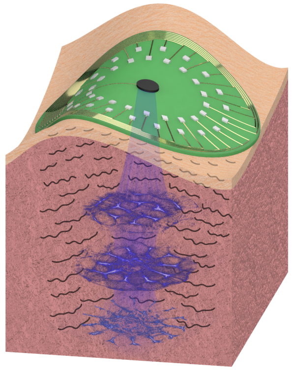 |
| Ultrasound sculpts spatial patterns of light in situ. |
In contrast, Chamanzar’s idea allows researchers to pattern and reprogram light as it propagates inside the brain (or any other tissue) without the need to insert implantable devices into the tissue. As a result, scientists will be able to mitigate the effects of scattering and other environmental influences. This cutting-edge, first-of-its-kind method of patterning light using non-invasive ultrasonic waves benefits from the best of ultrasound and optical
Maysam Chamanzar, who invented this novel technique and led the project, describes the idea: “Light is an electromagnetic wave, and ultrasound is a pressure wave. The interesting part of our idea is that we use waves to control waves. Both types of waves can propagate through the target tissue, for
This revolutionary approach to optical beam patterning improves the
 |
 |
 |
| Ultrasonically sculpted spatial patterns of light can be reconfigured in real time by changing the pattern of ultrasound. | ||
This method can be employed in several intriguing applications such as biological imaging, optical cancer therapy, and machine vision (such as seeing through foggy environments).
Chamanzar’s team published the results in Optics Express on Feb 27th, 2019 and is working to demonstrate that this technique can be used for functional recording and stimulation of brain activity in health and disease to understand and mitigate conditions such as epilepsy and Parkinson’s disease. Moreover, the team is now applying this technique to the field of optogenetics, as well as targeted non-invasive photodynamic therapy of cancerous
"This method has the potential to bring about a paradigm shift in the bio-photonic field. In fact, the biological tissue is usually an obstacle to light delivery in traditional optical methods. However, when ultrasonically modulated, the target medium itself can facilitate deeper penetration of photons," said Matteo Scopelliti, a graduate student in Chamanzar’s lab.
Yasin Karimi, another graduate student in the lab, explains the potentials: "Active control to shape and steer a complex pattern of
Perhaps even more exciting, their work is now being implemented in other fields as well. The publications have led to questions from peers who are now exploring its applications in robotics and machine vision, as well as in multi-point parallel imaging and
Searching for a minimally-invasive approach of interfacing with the brain, Chamanzar came up with the idea of using ultrasound to realize optical waveguides to guide light through the tissue non-invasively, while he was working on invasive neural implants at UC Berkeley. Chamanzar calls these invisible ultrasonically sculpted optical waveguides “virtual” waveguides. He performed proof-of-concept demonstrations of these virtual waveguides with Michel Maharbiz and Reza Alam (UC Berkeley), and Vikaas Sohal (UC San Francisco), which resulted in a continued collaboration among the teams after Chamanzar joined CMU and further developed this concept to demonstrate its utility for non-invasive light piping deep into turbid media such as brain tissue.
“The essence of optical waveguides such as optical
Regardless of how this research is fully realized, this innovative approach has the potential to drive a whole gamut of new applications in bio-photonics. With uses in a plethora of spheres, both medical and technological, Chamanzar’s work will have immediate and long-standing effects on different fields within science.