Insight without Incision: Advances in noninvasive brain imaging offers improvements to epilepsy surgery
About a third of epilepsy sufferers require treatment through surgery. To check for severe epilepsy, clinicians use a surgical procedure called electrocorticography (ECoG). An ECoG maps a section of brain tissue to help clinicians identify areas damaged by seizures.
“But ECoG requires taking a part of your skull out and putting electrodes directly on brain tissue,” ECE Professor Pulkit Grover said. An ECoG thus leaves a patient prone to infection. To find an alternative to ECoG, Grover’s team investigated making the non-invasive electroencephalogram (EEG) more effective by increasing electrode density and improving inference algorithms. He and the team recently presented their work at the annual meeting of the American Clinical Neurophysiology Society.
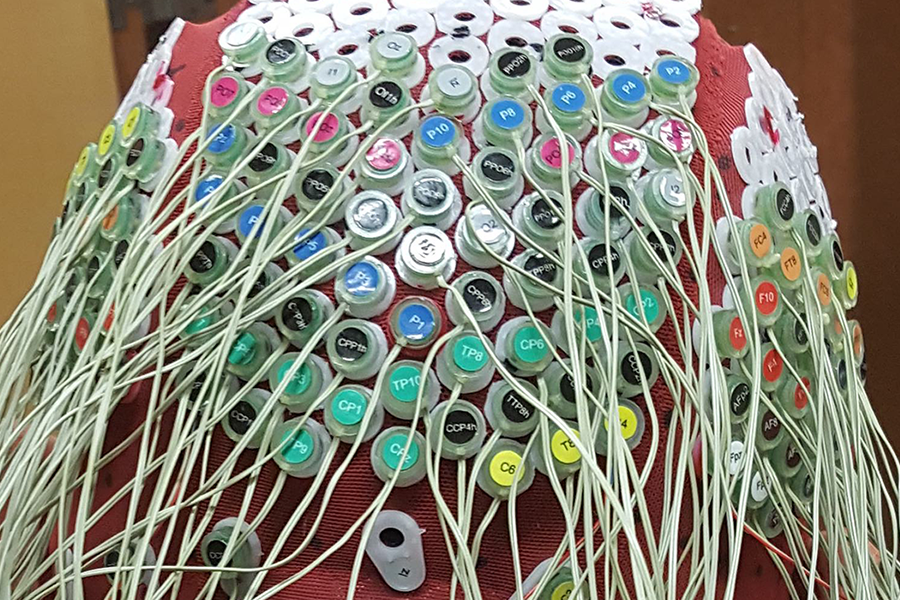
Treatment of epilepsy involves scanning the brain to locate the focus of epileptic seizures. A clinician administers an EEG scan by placing a cap with electrodes attached onto a patient’s head. The EEG records electrical function throughout the brain that the clinician then analyzes. EEG scans typically offer lower resolution imaging of brain activity than ECoG scans. This is a problem for EEG, since clinicians need to infer the location of focus of a seizure to perform the correct surgery. Grover’s study reveals that ECoG measurements do not carry the information of depth of focus, and hence its location, accurately enough to inform good surgical planning. This agnosticism can leave a patient’s illness inaccurately treated or, at worst, can reduce brain function in the operated area.
To improve EEG as a diagnostic tool, Grover, Ritesh Kumar, and Praveen Venkatesh worked on making EEG scans resolve deep brain activity with great fidelity and clarity. “The great surprise in this study,” Grover remarked, “is that the noninvasive Ultra-Resolution EEG outperforms ECoG in inferring depth of focus, even though ECoG sits much closer to the brain.” The Ultra-Resolution EEG modality was developed with Shawn Kelly, Marlene Behrmann, and Michael Tarr’s labs, and has been validated in separate experiments.
“One big limitation is that our current results are based purely on rigorous simulations with real brain and head models,” Grover emphasized, adding that his lab will pursue further experimentation with Dr. Mark Richardson at UPMC neurosurgery and Dr. Arun Antony at UPMC neurology, who also co-authored the study. Regardless, the study’s conclusions are promising. Grover and his lab have potentially discovered a way for clinicians to administer accurate and thorough care to epileptic patients without needing invasive diagnostics, or at least have provided better guidance for surgical inferences.
With Behrmann, Tarr, and Kelly at CMU, Grover is also investigating how Ultra-Resolution EEG could span other topics and fields, such as brain-machine interfaces and diagnosis of brain ailments from physical injuries to migraines.
CMU’s Rachel Sun and Gayathri Mohankumar also participated in the study. Venkatesh was supported by the Center for Machine Learning and Health fellowship. Early financial support to test the new EEG was provided by CMU's BrainHub initiative and ProSEED program. The work was also supported by an NSF WiFiUS award.