
| Overview | Cell Structures | Cell Migration | Cell Division |
Dependence of Cytokinesis on Midzone Microtubules in Multipolar and Nocodazole-Treated Cells |
Wheatley and Wang., J. Cell Biol. 135:981-989 (1996) |
The organization of midzone microtubules varies in cells that form an abnormal tripolar spindle. The correlation between the ingression pattern and midzone microtubules provides clues on how the two might be related. Microtubule dynamics in these cells were visualized by microinjection with rhodamine-labeled tubulin. Complementary experiments were carried out in cells treated with nocodazole after anaphase onset, where the incomplete destruction of midzone microtubules allows the analysis between ingression pattern and residual midzone structures. |
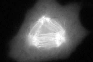 |
Division of a Cell with a Three-Segment Midzone into Three Daughter Cells |
Some tri-polar cells form midzone microtubules between each pair of spindle poles. These cells always ingress between each pair of poles and divide into three daughter cells. Recording time, 22 min. |
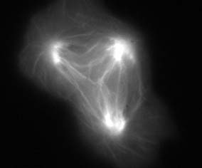 |
Division of a Cell with a Two-Segment Midzone into Two Daughter Cells |
Some tri-polar cells organize midzone microtubules between two of the three pairs of spindle poles. Typically one set of midzone microtubules move and merge with other midzone microtubules. Ingression takes place only between the poles with midzone microtubules, causing the cell to divide into two. In this cell no ingression occurs in the upper region. These observations suggest that the site of ingression is defined by the distribution of midzone microtubules. Recording time, 23 min. |
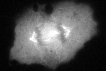 |
Stimulation of Ingression by Residual Midzone Microtubules in Nocodazole-Treated Cells |
Cells treated with nocodazole during anaphase typically show only partial disruption of midzone microtubules. Cortical ingression in these cells shows an irregular path, "chasing" the residual microtubule bundles. This adds evidence that the site of ingression is defined by midzone microtubule bundles. Recording time, 67 min. |